Modern medical use of computerised tomography (CT) is increasing. On this page we explore the risk of cancer associated with CT scanning, which may be in the region of 1:4000, or even more severe. Radiation from a single CT scan is equivalent to about 100 to 250 plain chest films, and is showing no signs of decreasing with modern CT scanners. Many physicians seem unaware of this radiation dose, and the associated risks. (Lee et al, 2004). It would seem wise to factor such risks into decision making about whether or not to perform a CT. We should also inform patients of risk, particularly where a young person is having a CT.
We will in turn discuss terminology, the radiation risks of CT scanning, data from A-bomb exposure, and counter-arguments. We will then present a practical approach followed by a brief look at the future.Patients are more likely to sue their doctor (and win) if the doctor doesn't adequately tell them of the risks of the procedure they're undergoing. This is graphically shown in the Australian case of Rogers versus Whitaker, where the surgeon failed to caution the patient about the 1:14 000 risk of surgery to a damaged eye. When the patient subsequently developed near-total loss of vision due to sympathetic ophthalmia, the damages awarded were substantial, not related to the competence of the surgery, but because of poor communication of risk.
Radiation terminology is made more confusing because there are old and new-style units. One of the merits of the older units --- roentgens and rads and rems --- is that one Roentgen of x-rays produces about a rad of tissue dose of radiation, which has a rem of biological effect. So for x-rays at least, we could say with some confidence that `a Roentgen is a rad is a rem'!
In the new system, Roentgens, rems and rads have all been replaced by new, unfamiliar units. These measures, all bigger than the convenient older units, are summarised in Table 1, together with the Becquerel and curie, which are simply counts of the number of radioactive disintegrations of atoms. The biological effect of the radiation depends not only on the amount of radiation, but also on the type of radiation being emitted, what tissue is being irradiated, and how much of the radiation is being absorbed. One can `guesstimate' the number of Sieverts from the number of Grays. For a lot of the radiation we deal with, notably gamma and x-rays the conversion factor is simply 1. This coefficient we multiply by is termed the RBE or `relative biological effectiveness'. Current values are specified in the ICRP `Radiation Weighting Factors' as 5--20 for neutrons (depending on energy), 5 for protons, 20 for alpha particles, and 1 for x-rays, gamma rays and electrons.
| Table 1 | |||
| New Measure | Old unit | Conversion | Interpretation |
| Becquerel (Bq) | curie (Ci) | 1 Ci = 37 billion Bq | Disintegrations per second. Of little immediate biological usefulness! |
| Coulombs per kg (C/kg) | Roentgen (R) | 1 C/kg = 3876 R | The Roentgen was originally the amount of electromagnetic radiation producing an `electrostatic unit of charge' (esu)* in a cc of dry air at 0 o C and 1 atmosphere of pressure! |
| Gray (Gy) | rad | 1 Gy = 100 rad | The Gray (Gy) is a measure of absorbed dose, the older unit being the rad , an acronym for radiation absorbed dose . There are 100 rads in one Gray. A Gray represents the transfer of 1 Joule of energy per kilogram. (That's a lot of radiation!) |
| Sievert (Sv) | rem | 1 Sv = 100 rem | The scaling factor of 100 is the same as for Gray/rad. The term `rem' is an acronym for `Roentgen equivalent man', a clear reference to the rem being an indication of radiation effect on biological tissue. |
| *An esu is the unit of electrical charge used in the cgs system of units. It's about a third of a billionth of a Coulomb. It's also called the Franklin or `statcoulomb' | |||
Most authorities quote similar risks for exposure to 10 mSv of radiation: a long-term cancer risk of about 1:2000. This is the value quoted by the United states Food & Drug administration (FDA, 2005), while the National Council on Radiation Protection and Measurements (NCRP) estimates the lifetime risk of cancer for the same dose at 1:2500, as does the Health Physics Society. All of these authorities appear to base their risk assessment on a committee called `BEIR V', an acronym for Biological Effects of Ionizing Radiation.
There's some imprecision. Here's a quote derived from the executive summary of BEIR V:
"Due to sampling variation alone, the 90% confidence limits for the Committee's preferred risk models, of increased cancer mortality due to an acute whole body dose of 0.1 Sv to 100,000 males of all ages range from about 500 to 1200 (mean 760); for 100,000 females of all ages, from about 600 to 1200 (mean 810)."
In addition, there are other cautions:
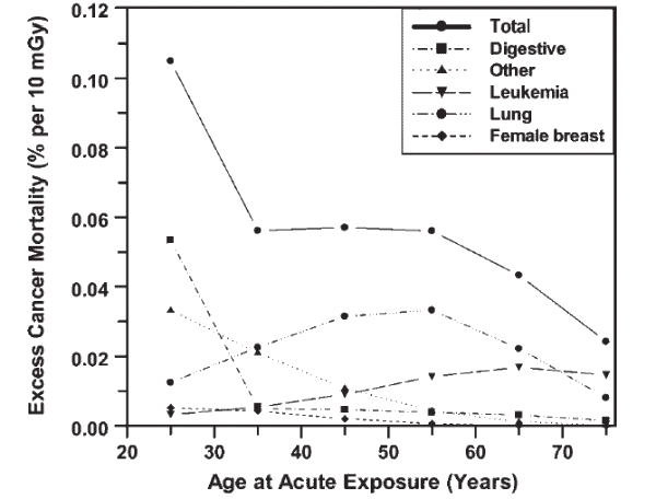
Here's a graph from BEIR V (See Brenner & Elliston, 2004) which shows how total risk of cancer goes up more at lower age, with 10 mSv (Note label says mGy) radiation dose.

Although recent (2000-2002) estimates suggest that only about 13% of radiology procedures involve CT, these procedures are said to provide about 70% of all in-hospital radiation (Lee et al, 2004). The dose from a chest CT is equivalent to about one hundred to two hundred and fifty plain chest films.
Approximate dose values (in milliSieverts) for various procedures, as adapted by the FDA from EC data are:
| Diagnostic Procedure | Typical Effective Dose (mSv) |
| Chest x ray (PA film) | 0.02 |
| Skull x ray | 0.07 |
| Lumbar spine | 1.3 |
| I.V. urogram | 2.5 |
| Upper G.I. exam | 3.0 |
| Barium enema | 7.0 |
| CT head | 2.0 |
| CT abdomen | 10.0 |
Browsing through the literature, you'll often find values in the range of about 4--7 mSv for chest CT scanning, and about 6--20 mSv for abdominal CT. Some report higher values. There has been no decline in radiation dose from CT in the past ten years, in fact, if one takes slice overlap into account, the dose with faster new machines may even have gone up. A recent study of multi-detector row CT versus single detector CT showed a 27% increase in radiation dose. Others have reported even higher dose increments.
There seems to be some good news, however. Recent simulations (Tack et al, 2005) suggest that diagnostic quality of images isn't significantly degraded in some circumstances (CT pulmonary angiography) despite substantial reductions in radiation dose (as measured by the `tube current-time product').
You would think that we could get a lot of information about radiation risk from the surviving victims of A-bomb blasts. We can, but here too there has been vast, recent controversy about how much radiation was inflicted upon survivors of Hiroshima and Nagasaki. (The radiation here was predominantly neutrons and gamma rays, so we may not be able to extrapolate directly to other forms of radiation exposure, but the Sievert as a measure of radiation effect still comes to our aid).
There is a substantial body of information about cancer incidence in A-bomb survivors who received lower doses of radiation. Despite whinging from both right and left, the picture seems to be one of linear dose response. Here's a graph from RERF, which in less politically correct times was called the `Atomic Bomb Casualty Commission', (RERF, 2001):
With any hot topic, there's a wide distribution of opinion, none more so, perhaps, than when we're dealing with radiation. On the extreme `right' we seem to have the belief that radiation (and especially nuclear power) is the solution to all ills; on the far `left' are those who believe that even minuscule doses of radiation are hundreds of times more likely to cause cancer than the estimates given by, for example, BEIR V.
Fortunately for us, the fanatics on either side usually betray their bias. For example the radioactive right tend to diminish incidents such as Three Mile Island as `valuable learning experiences'. They selectively read the UNSCEAR report on Chernobyl, often briefly dismissing the massive rise in thyroid cancer in affected areas, and concentrating on the current absence of evidence of other carcinogenic effects (yet). They enthuse about potential benefits (hormesis) of low-dose radiation on the immune system.
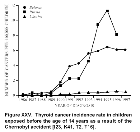
As an aside, let's look at Chernobyl, where nine tons of radioactive material was released into the atmosphere. Much of the radiation released by the meltdown was in the form of radioactive iodine, which is avidly taken up by the thyroid gland, so we would expect a lot of our cancers to be thyroid-related. After Chernobyl, thyroid doses were estimated at "at most 25 mGy for one-year old infants". People in Belarus copped a lot of the dose due to wind direction, and much of the Ukraine was spared. Here's a graph from UNSCEAR (UNSCEAR, 2000):

I find this graph quite chilling; if people dismiss it lightly, I'm afraid I tend to do the same with their glowing reports of the benefits of radiation --- hormesis and whatnot.
On the other side we have those I term the `dangerously low left' --- those who maintain that even minuscule added doses of radiation vastly increase the risk of cancer. These assertions seem to be at variance with the effects of background radiation, which as we note below, is several millisieverts, even at sea level. If low levels of radiation were that damaging, surely we'd be dropping like flies, or, come to think of it, almost exactly not like flies, which are fairly radiation-resistant!
The truth may well lie somewhere in between. I prefer to regard the two extremes as populated by `antiparticles' which if brought together would likely result in mutual annihilation (Probably in the process producing large amounts of damaging hard radiation).
I believe we need a sense of perspective. The evidence seems to favour adopting the middle ground. We mentioned `a walk in the park' in our title, but even such a stroll is not without risk! At sea level, despite the fact that the atmosphere above us provides some protection against radiation, we are still exposed to about 2.6 mSv per year, some arising from energetic particles produced during nuclear processes in the sun, and other from background cosmic radiation. At an altitude of 3200 m this irradiation may increase to 12.5 mSv. So you will be naturally irradiated, even walking in the park, and the dose depends very much on where you are living. In some areas even the rocks (particularly granite) can exposure you to 20 mSv per year, or more!
This background radiation (with a probable attendant risk of cancer) is no reason for complacency. The bulk of evidence suggests that radiation in the range of 5 to 10 mSv produces a significant increase in the risk of cancer. A single CT scan can provide more radiation than you'd otherwise receive in a year.
(Interestingly enough, the NCRP recommends an annual radiation dose limit for individual members of the public from all radiation sources other than natural background and the individual's medical care as 1 mSv (NCRP, 2004). Infrequently in that individual's lifetime, they permit a dose up to 5 mSv. There's a lot of fine print, and radiation workers are strangely exempt. Even more peculiar is the failure to classify airline flight crews as `radiation workers', despite the several extra millisieverts they receive every year!)
CT scans are often valuable investigations, and sometimes they are simply the best choice. But I believe that when we perform them we should:
In older individuals, the tiny risk of cancer from a CT will usually be drowned out by the high background risk of cancer in the aged. About one in five people will ultimately die of cancer. In addition, older people will have less time in which to develop cancer due to radiation exposure. Not so in the young!
For example, in a young person with a normal chest film and a fairly low a-priori risk of pulmonary embolism, rather than performing a CT pulmonary angiogram, a perfusion scan may well do the trick at one tenth of the radiation dose.
In order to explore the future of medicine, litigation and radiation damage, we need to digress briefly and look at mechanisms of mutation. Here goes ... (We assume you know a little about how DNA works)!
The important message is that different mutagens (things inducing mutations in DNA) work in different ways, and spontaneous mutations also commonly occur. The following section is a little technical, so if you wish you can skip to the main point.
Various changes can occur in DNA bases, including transitions (purine is replaced by another purine, pyrimidine is replaced by another pyrimidine) or transversions (pyrimidine/purine replacement), deletion of bases or insertion of new ones, and in extreme cases breaks and rejoining of whole DNA strands. Let's explore some such changes:
DNA methylation is a normal mechanism for both protecting DNA and suppressing transcription. However methylcytosine can be deaminated to thymine, which is a normal component and cannot be detected and repaired. Particularly prone to such deamination are genes containing the dinucleotide CG. There are two consequences of this quirk of DNA: firstly, the CG sequence is about five times less common than one would expect, and secondly, the body prevents methylation of CG sequences in gene exons. In fact, the presence of hypomethylated CG (CpG islands) suggests that a gene is present, and that the DNA is not simply 'junk' --- this is how the cystic fibrosis gene was first identified! Mutation `hot-spots' occur near CpG islands, for example in the gene coding for achondroplasia!
Another mechanism of spontaneous point mutation occurs because bases in DNA can shift between two forms (isomers) called `keto' and `enol' forms. This shift is called tautomerisation. For example, G is normally in the keto form and therefore pairs with C, but in the enol form G will instead pair with T instead!
The normal cell has powerful defences against many of the above mutations, so DNA transcription is phenomenally accurate. (Proofreading mechanisms have been extensively studied in Escherichia coli, for example, where the error rate may be as low as 1:10 11 base pairs!) Apart from the defences already discussed, a potent defence is the 3'-5' exonuclease which `proofreads' and removes incorrectly paired bases. DNA ligase I rapidly repairs single-strand DNA breaks. There are several other specific glycosylases apart from the uracil glycosylase mentioned above.
Nucleotide excision repair involves detection of a distorted double helix and excision of the damaged region (defective in xeroderma pigmentosum, explaining the sensitivity to sunlight, as the UV-induced dimers aren't clipped out)! As there is still a residual loss of information, the importance of this repair mechanism may be that it fixes things enough to allow continued DNA replication. (Such mutations and their repair may explain the enormous amount of `junk' DNA kicking around in our genetic material --- perhaps it's there to take the hits).
Most important of all in the defence against mutation may be the presence of 'checkpoints' in the cell cycle. If the DNA has been grievously damaged, it's likely that the cell will commit ritual suicide (apoptosis) rather than proliferating. Defects of this checking mechanism (as are seen in ataxia telangiectasia and the Li Fraumeni syndrome) result in poorly controlled proliferation of defective cells, and ultimately in cancer.
You can see that although gene mutation and the body's defence against such damage is hellishly complex, different mutagens do different things. It's conceivable that in the future, we may be able to look at an individual (together with their cancer) and decide, based on probability, what caused that cancer. We may even be able, by looking at their genome and how it's been damaged, to step back through the processes which caused their cancer. Adjacent, more subtly damaged cells may give us further clues about damage mechanisms.
If someone's cancer is likely to be radiation-induced, and they have been irradiated by their doctor without caution and a discussion of risk/benefit ratios, this may be fertile ground for litigation. The numbers are there (as stated by the FDA and NCRP, who may take precedence over gainsayers on both left and right, at least in the eyes of the law) --- what we need to do is pass on the message. More important still is simply not to expose your patient to unnecessary risk!
 http://www.unscear.org/pdffiles/annexj.pdf.
The full report is
  here.
here.
 http://www.ncrponline.org/NCRP%20Statement%20No.%2010.pdf
Apart from the above references, I've inserted links and article references throughout the HTML source code for the above document (This is to make the page less densely packed with relatively minor links). Click on View|Source in your browser if you're interested.
| Date of First Publication: 2005/08/26 | Date of Last Update: 2006/10/24 | Web page author: Click here |