Results
Fifty-eight patients who died after sepsis underwent autopsies. Autopsy
revealed that the spleen was enlarged several times in size and its weight
occasionally reached 900 g (mean weight 305+/-115 g). Microscopically,
the splenic sinuses were engorged with blood and contained many polymorphonuclear
leukocytes and occasionally bacteria. There was marked fatty and parenchymatous
degeneration of heart, liver, and kidney. Myeloid hyperplasia was the most
common finding in bone marrow. Lymph nodes were increased in size. Microscopically,
the enlarged lymph nodes contained immunoblasts and plasma cells. In most
of the DIC syndrome cases, besides signs of numerous petechial hemorrhages
and alterations in small veins that varied from endotheliosis to septic
trombophlebitis. Variants of septic process development are listed in Table
2.
| Table 1. Variants of Sepsis |
| Variant |
Number of Patients(%) |
| Otogenic |
2(2.5) |
| Odontogenic |
3(3.7) |
| Gynaecological |
6(7.4) |
| Urological |
11(13.6) |
| Following IV cannulation |
11(13.6) |
| Post-surgical sepsis |
28(34.5) |
| Cryptogenic |
20(24.7) |
As seen in Table 1, amongst the diseases, the
leading cause of death was sepsis related to surgery. Septic patients died
mainly from overwhelming sepsis, multiple organ failure (63.8%)
and encephalopathy(19%).
The etiologic agents of sepsis were identified in only 14 of 81 patients
as seen in Table 2.
| Table 2.
Pathogens isolated from the blood of septic patients |
| Pathogens
Gram-positive aerobes |
Number of Patients (%) |
| Staphylococcus aureus |
7(8.6) |
| Streptococcus pyogenes |
1(1.2) |
| Streptococcus viridans |
1(1.2) |
| Gram-negative aerobes |
|
| Enterobacter |
1(1.2) |
| Pseudomonas aeruginosa |
1(1.2) |
| Acinetobacter |
1(1.2) |
| Klebsiella |
1(1.2) |
| Citrobacter |
1(1.2) |
Demographic analysis did not reveal any statistically significant
differences in age, gender, duration of sepsis, prehospitalisation period,
frequency of septic shock episodes and occurrence of MOF in both groups.
( Table 3) This suggests that the two groups are
comparable.
|
Table 3. Demographic characteristics of patients with sepsis.
|
|
Group 1
n=23 |
Group 2
n=58 |
| M/ F |
13/10 |
22/36 |
| Median age
(range) |
35.2
(16-66) |
50.2
(17-84) |
| Sepsis duration before hospitalisation
(days) |
13.1
(1-60) |
22.3
(1-150) |
At the same time there was a significant difference in frequency
of application of EDM between survivor and nonsurvivors groups. The analysis
revealed that EDM was used significantly more often in patients that survived
sepsis (Group 1). Thus in Group 1, ChH was used 3 times more frequently,
UBI 8 times more frequently, PSC 10 times more frequently and HBO 4 times
more frequently than in patients that died (Group 2)(Figure 1). We hypothesized
that combination of EDM and HBO influenced on mortality rate in
septic patients. In order to evaluate this hypothesis all 81 patients were
reallocated in one of two new groups according to the principle of application
of EDM and HBO. Group 3 - 40 patients which have been treated with traditional
therapy and group 4 - 41 patients in which traditional therapy was supplemented
with EDM and HBO. Demographic characteristics of patients 3 and 4 groups
are summarized in the Table 4.
|
Table 4. Demographic characteristics of patients with sepsis.
|
|
Group 3
n=40 |
Group 4
n=41 |
| M/ F |
14/26 |
23/18 |
| Median age
(range) |
51,2
(21-84) |
41,0
(16-77) |
| Sepsis duration prior to hospitalisation
(days) |
25.6
(1-80) |
15,6
(1-150) |
The demographic analysis did not reveal any significant statistical
differences in age, gender and duration of the period before hospitalization
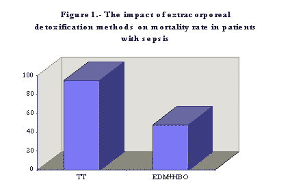
in both groups. Nevertheless in group 3 mortality
rate was 95%, in contrast to a mortality rate of 55% in group 4 (p<0.05
) ( Figure 1).
TT = traditional therapy
EDM+HBO = extracorporeal detoxification + hyperbaric oxygen
After starting the extracorporeal detoxification program, the biochemical
variables reflecting MODS (bilirubin, hepatic enzymes, creatinine and
MMWC) came close to normal values within 14 days.
Discussion
The occurrence of liver failure and renal failure
in patients with sepsis is thought to be due to entry into the circulation
of toxic metabolites (creatinine, urea, neurotoxins, bilirubin,
bile acids, middle molecular weight compounds, etc.) that are able, by
themselves, to stimulate the syndrome of endogenous intoxication. Aside
from this disorders in microcirculation distinctly reduce the
rate of elimination of toxic metabolites (18)
Since Yatzidis and colleagues (19) published their individual reports,
ChH has become increasingly popular with surgeons and specialists in hemodialysis.
During the past two decades the great numbers of reports of the effective
usage of ChH, in patients with exogenous and endogenous
'endotoxicoses' have been
reported in the CIS (USSR). Mainly they are summarized in several monographs(8-10,20).
Apart from classic hepatic toxins - ammonia, free fatty acids, mercaptan
and phenol - carbonaceous sorbents remove other toxic
metabolites from the circulation, including creatinine, guanidinoacetic acid,
and bilirubin (10,21-24).
Non-coated charcoal sorbents are able to absorb on their surface such
substrates of endogenous intoxication as middle molecular weight compounds
(MMWC)(25-27). We assert the importance of measuring the concentration
of MMWC in serum as a leading sign of endogenous intoxication. The four
-fold increase of MMWC in serum is by itself grounds for starting EDM.
Normalization of MMWC in serum practically always is associated with the
regression of MODS. In the 1980s the generally accepted idea about ChH
was that a pure absorption process has occurred. It was demonstrated that
autotransfusing blood after its contacting with sorbent was able to alter
the sorption activity of the glycocalyx of all circulating erythrocytes.
This phenomenon was combined with amelioration of peripheral tissue perfusion
(28). The other nonabsorbtion phenomenon of ChH was that it prolonged the
remission and enhanced the sensitivity to basic medicines in patients with
autoimmune diseases (29-30).
Currently ChH is a method of treating the toxic syndrome seen in patients with peritonitis,
necrotizing pancreatitis, gangrene, and other purulent surgical diseases
(10,31,32)
The earliest reports on PSC as a method of immunomodulation in septic
patients are from 1985 to 1986 (33-35). It is presumed that the grounds
for applying PSC in animal models and clinical trials were the observations
that asplenic patients or those who underwent splenectomy were prone to
overwhelming infection or fatal sepsis which was sometimes associated
with DIC (36-39). Septic complications in asplenic patients were explained
by the decrease in antibody production(40) and disorders in tuftsin synthesis
(41), but still remain poorly delineated. In 1885 Shumakov et al., for
the first time demonstrated that the donor's (pig) spleen allo- and xenograft
when connected extracorporeally makes the leukocyte phagocytes active in
septic animals and patients (PSC has been used in 20 patients with sepsis
(34)). Grinev et al, in 1986 applied PSC in 14 patients with mechanical
trauma and subsequent septic complications. The authors observed twice
as many blood leukocytes in the 24 hours immediately following
the procedures (37). It
was demonstrated that PSC normalized the activity of natural killer cells
that had been depressed considerably before xenoperfusion (42). Moskalenko
et al. report showing positive effects by using PSC in 14 patients with
purulent septic diseases (43). Ryzhalo et al. applied PSC in the combined
treatment of surgical burn sepsis in 5 patients(44). Sitnikov et al. simplified
the PSC method. In 1992 they reported the results of using hemoperfusion
through the porcine spleen followed by the infusion of xenosplenic perfusate
in the treatment of purulent peritonitis and various suppurative complications
in 109 patients. A positive effects resulted in 20% reduction mortality
rate with 1.5-3.5 times shorter period of treatment (45).
UBI proved to be effective in the treatment of purulent diseases of
the maxillofacial region (46) and severe sepsis when combined with ChH
(47). Many studies done on the effects of UBI concerning immune and nonspecific
responses have shown them stimulated after a few UBI procedures. It has
been demonstrated that UBI increased the number of monocytes and lysozyme
concentration in blood, raised the index of T-lymphocytes blast transformation
(48) and improved some rheological characteristics of blood. This allowed
it to be recommended as a supplemental method in patients with ischemic
heart disease (49).
The use of HBO in intensive care is a well-established procedure used
for many years. The reported advantages of HBO in critically ill patients
include reductions in mortality of 95% in patients with acute myocardial
infarction (50), the normalizing of PaO2, and a decrease in serum creatinine,
billirubin and ammonia level in patients with acute renal failure (51).
It has been reported that HBO is conducive to the repair of reversible
(glutamine synthesis) and irreversible (urea synthesis) routes of ammonium
binding in hepatocytes. These are disordered in chronic hepatitis (52).
HBO when combined with ChH improved the functional capacity of cardiac
muscle, facilitated reversal of anaerobic metabolism and
normalized oxygen consumption (53).
Thus the extensive literature on EDM provides evidence that EDM
decreases the level of toxic metabolites in blood and is able to temporarily
support body detoxification systems. The present study demonstrates
that EDM was used significantly more often in survivors than in the patients
who died after sepsis. In patients treated with the combination of traditional
methods plus EDM, the mortality rate was half that of patients treated with
traditional methods.
The beneficial effects of applying EDM in septic patients may be related
to two interacting mechanisms. First, there is the effect of detoxification
due to the removal of toxic metabolites from circulation. Second, there
is a systemic effect linked with the augmentation of the host defense system
that is compromised in sepsis, as our previous studies and other author's
studies have shown (34,35,42). The combined use of EDM in this study to
a certain extent reflects the evolution of our understanding
of sepsis. Sepsis is not only a state in which the defences of the host
are compromised but when combined with MOF, it is also a consequence of
accumulation of endogenous toxins.
(* = References not found in Medline)
-
Spengler RF.,Greenough WB III,Stolley PD: A descriptive study of nosocomial
bacteremias at the Johns Hopkins hospital,1968-1974.Johns Med J 1978;142: 77-84.
-
Bryan CS,Reynolds KL,Brenner ER: Analysis of 1,186 episodes of gram-negative
bacteremia in non-university hospitals; the effects of antimicrobial therapy.
Rev Infect Dis 1983;5: 629-638.
- *
Sepsis. in : Big Medical Encyclopaedia. Sovetskaja encyclopaedia(Ed).Moskva,1984;23: 325-326.
- *
Waage A,Espevik T,Haistensen A: TNF,IL-1,and IL-6 in human septic shock
. Scand J Immunol 1988;28: 267-271.
-
Sullivan JS,Kilpatrick L,Costarino AT,et al: Correlation of plasma cytokine
elevations with mortality rate in children with sepsis. J Pediatr 1992;120: 510-515.
-
Fisher CJ,Opal SM,Dhainaut JF,et al: Influence of an Anti-Tumor necrosis
factor monoclonal antibody on cytokine levels in patients with sepsis.
Crit Care Med 1993;21: 318-327.
-
Pinsky MR,Vincent JL,Deviere J,et al: Serum cytokine levels in human septic
shock: relation to multiple-system organ failure and mortality. Chest 1993;103: 565-575.
- *
Nikolaev VG, Strelko VV: Hemosorption on activated charcoals. Naukova Dumka
(Ed). Kiev,1979,286 (Rus)
- *
Nikolaev VG: Method of hemocarboperfusion in experiment and clinic. Naukova
Dumka(Ed).Kiev,1984,301 (Rus)
- *
Lopatkin NA,Lopuhin YrM: Efferent methods in medicine.Medicine(Ed). Moskva,1989,351
(Rus)
-
Bone RC: Let,s agree on terminology: Definitions jf sepsis. Crit Care Med
1991;19: 973-976.
-
The ACCP/SCCM Consensuns conference commitee: definitions for sepsis and
organ failure and guidelines for the use of innovative therapies in sepsis.Chest
1992;101: 1644-1655.
-
Fry DE,Pearlstein L,Fulton RL,et al: Multiple system organ failure: The
role of uncontrolled infection. Arch Surg 1980;115: 136-140.
-
Knaus WA,Draper EA,Wagner DP,et al: Prognosis in acute organ system failure.
Ann Surg 1985;202: 685-693.
-
Koch KA,Rodefer HD,Wears RL: Changing patterns of terminal care management
in an intensive care unit.Crit Care Med 1994;22: 233-243.
-
Frankenfield DC, Reynolds HN, Wiles III ChE,et al: Urea removal during
continuous hemodiafiltration. Crit Care Med 1994;22: 407-412.
- *
Knott EK: Development of ultraviolet blood irradiation. Amer J Surg 1948;76: 165-171.
- *
Umansky MA, Pinchuk LB, Pinchuk BG: Syndrom of endogenous intoxication.
Naukova Dumka (Ed).Kiev,1979,201 ( Rus)
- *
Yatzidis H: A convenient hemoperfusion micro-apparatus over charcoal for
the treatment of endogenous intoxication. Its use as an effective artificial
kidney.-Proc Eur Dial and Trans Ass 1964;1: 83-86.
-
Nikolaev VG,et al: Early experimental studies on hemoperfusion as a treatment
modality for acute radiation disease.Artif Organs 1993;17: 362-365.
-
Chang TMS: assessments of clinical hemoperfusion in uremic patients. Clin
Nephrol 1979;11: 111-119.
- *
Chang TMS: Sorbents and their clinical application.New York,1980,217.
-
Chen CZ,et al: Clinical trials for removal of bilirubin by high-capacity
nonionic adsorbent.Artif Organs 1993;17: 76-78.
-
Riabtsev VG,Solomka YaA: Combined treatment of mechanical Jaundice. Khirurgia
1994;5: 38-42 (Rus).
- *
Simbirtsev SA,Belyakov NA: Pulmonary microembolism.Medicina(Ed). Leningrad,1986,216
(Rus).
-
Kanus II,Ilukevich GV,Kljavsunnik IZ: Effectiveness of hemosorption in intensive
care of patients with Lyell's syndrome. Anesteziol Reanimatol 1993;6: 63-65.
-
Gutnikova AR,Alimov MM,Abidova SS,et al: Change in the concentration of
nontoxic plasma components in the process of hemosorption in experimental acute
liver failure.Patol Fiziol Eksp Ter 1994;1: 30-32 (Rus).
- *
Zhidkov CP: Hemoperfusion methods as a variant of extracorporeal therapy
of peptic ulcer.Abstract of thesis Doc Med Sci.Sanct-Petersburg 1991,22.
- *
Shumakov VI,Dmitriev AA: Promising results results with hemoperfusion in
patients with immunorelated diseases.Past,Present and Future of artificial
organs.Piskin E(Ed).Ankara,1983: 225-228 (Rus).
- *
Shumakov(Ed):
Artificial Organs.Medicina.Moskva,1990,205.
-
Ternovoy KS,Bulakh AD,Kosyakov AN: Complex sorption therapy of locomotor system
suppurative complications.Biomater Artif Cells Artif Organs 1987;15: 31-39.
-
Genyk SN,Grushetsky NN: Clinical course of purulent and necrotic complications in diabetes mellitus. Khirurgiia
1993;5: 28-31 (Rus).
-
Shumakov VI,Tsypin AB,Safarov Syu,et al: Extracorporeal connection of donor's
spleen for detoxification of the organism.Khirurgia 1985;4: 110-114 (Rus).
-
Grinev MV,Tsypin AB,Tarelkina MN:
An experience with the extracorporeal connection of the
xenospleen in patients with traumatic shock. Vestn Chir 1986;11: 81-86
(Rus).
-
Zhidkov KP,Medvedev YuA,Dobrynsky EK: Extracorporeal perfusion of the xenospleen
in the treatment of purulent surgical diseases.Vestn Chir 1986;11: 86-91
(Rus).
-
Eraklis AJ,Kevy SV,Diamond LK,et al: Hazards of overwelming infection after
splenectomy in childhood.New Engl J Med 1967;276: 1225-1229.
- *
Quie PG,Davis AT: Phagocytic and granulocytic disorders. in : Immunologic
disorders in infants and children.Stienhm ER,Fulgimiti VA(Eds). Philadelphia,1973,273-288.
-
Likhite VV: Immunological impairment and susceptibility to infection after
splenectomy.JAMA 1976;236: 1376-1377.
-
Boughton BJ,Simpson A,Chandler S: Functional hyposplenism during pneumococcal
septicemia.Lancet 1983;8316: 121-122.
-
Lozzio BB,Wargon LB: Immune competence of hereditarily asplenic mice. Immunol
1974;27: 167-178.
-
Costantopoulos A,Najjar VA,Smith JW: Tuftsin deficiency: a new syndrome with
defective phagocytosis.J Pediat 1972;80: 564-572.
-
Zhidkov KP,Malygin AM,Dmitriev NV,et al: Changes in natural killer activity
of peripheral blood lymphocytes in patients with pyoseptic diseases under
the effect of extracorporeal perfusion of pig spleen sections.Anesteziol Reanimatol
1989;1: 37-40 (Rus).
-
Moskalenko VZ,Fuxzon AS,Zhurilo IP,et al: Hemoperfusion with the use of
porcine spleen in the treatment of purulent -septic disesases in children.
Klin Khirurgia 1992;6: 24-26 (Rus).
-
Ryzhalo MN,Shumakov HSh,Sidelnikov VO: Hemoperfusion via a xenogenic spleen
in the combined treatment of surgical burn sepsis. Voen Med Zh 1992;8: 26-27
(Rus).
-
Sitnikov VA,Cipin AB.Stjazhkina SN,et al: The antitoxic action of xenogenic
spleen in treating the supurative septic complication in surgery. Vestn
Khir 1992;148: 101-105 (Rus)
- *
Dmitrtieva VC,Alexeeva AN,Veretenik GI,et al: Usage of ultraviolet blood
irradiation at inflamatory process in maxillofacial region. Vestn Khir
1983;6: 119-122.
- *
Karjakin AM,Kucher VV,Susla PA,et al: Hemoperfusion and ultraviolet blood
irradiation in treatment of severe sepsis. Vestn Khir 1983;4: 109-112.
-
Ganelina IE,Samoilova KA: Mechanisms of influence of ultraviolet blood irradiation
on humans and animals organizm.Nauka(Ed),Leningrad, 1986,264.
-
Ganelina IE,Kukui LM,Nikolaewa,et al: Zur tharapie schwerer stenikardien
mittels ultraviolet-bestrahlung des blutes(UVB) und zu einigen wirkungsmechanismen
dieser therapie.Folia haematol 1982;109: 470-482.
-
Gonchar DI: Hyperbaric oxygenation in myocardial infarction. Anesteziol
Reanimatol 1994;3: 54-56. (Rus)
- *
Efuni SN: Handbook of hyperbarooxygenation.Medicina(Ed).Moskva,1986,415.
-
Savilov PN,Leonov AN,Yakovlev VN: Role of hyperbaric oxygenation in the
mechanism of ammonium detoxification in resection of the liver in the presence
of chronic hepatites. Anesteziol Reanimatol 1994;6: 31-34.
-
Rodionov VN: The effect of hemosorption and hyperbaric oxygenation on the
indices of systemic hemodynamics and oxygen allowance in patients with
septic shock. Anesteziol Reanimatol 1994;2: 45-47.