Manage wide-complex tachycardia
Follow the decision tree.Step 0: Is the patient haemodynamically unstable?
Comment: Haemodynamic instability means that the arrhythmia should be addressed immediately. The following algorithm cannot be used where the ventricular response rate is irregular. As an aside, note that one should also include artefact in the differential diagnosis (on a rhythm strip alone), as shown in Knight's recent paper!References:
|
Step 1: Is any one of the following present: ischaemic heart disease, or heart failure, or past heart surgery, or cardiac enlargement?
Comment: One can argue that the a priori likelihood of VT is so high in patients with previous myocardial infarction (or substantial organic heart disease) that one should manage as for ventricular tachycardia without wasting further time evaluating the ECG! (See the article by Gupta & Thakur, or that of Shah et al).References:
|
Step 2: Are there any RS complexes in the V leads?
![]()
Comment: This and the following steps are based on Brugada's algorithm for diagnosis of wide-complex tachycardia. We have slightly modified the approach to make it more workable. Please consult the excellent original reference. Absence of any RS complex in the precordial leads V1..V6 is 100% specific for VT, but is insensitive (26%). Isenhour et al recently re-evaluated Brugada's algorithm, - their four doctors were less effective in differentiating VT and SVT than in the original paper, perhaps because the later study included some patients on drug treatment for arrhythmias (excluded from Brugada's study).References:
|
Step 3: In any of these RS complexes, is the interval
from start of R to nadir of S > 100ms ?
![]()
Comment: This criterion of Brugada is based on the initial observation by Kindwall et al. (whose criteria differ somewhat from those of Brugada in that they used 60ms as the cutoff, and only looked at V1 and V2). A duration of over 100ms almost excludes SVT (98% specific for VT).References:
|
Step 4: Is there AV dissociation,
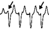
or are there fusion or capture beats?
{A capture beat QRS looks normal because a conducted sinus beat transiently captures
the ventricles;
a fusion beat's QRS is intermediate
between normal and the VT complex}
Comment: These are traditional criteria for VT (and pretty reliable when present). In Brugada's study, evidence of AV dissociation was 98% specific for VT. References:
|
Step 5: Is the QRS complex in V1 predominantly positive?
![]()
Comment: More 'conventional' criteria. There is a wide variety of other criteria that have been proposed, for example 'concordance' (similar QRS in V1 through V6), left axis deviation, a QRS duration over 140ms, and so on. Many such criteria have been found wanting, as discussed by Brugada in his 1991 paper! |
V1 predominant positive:
Look at V1. Is there?
a monophasic R
![]() or a QR
or a QR
![]() or an RS
or an RS
![]()
a monophasic R
![]() or a QR
or a QR
![]() or an R:S ratio < 1
or an R:S ratio < 1
![]() or a QS
or a QS
![]()
V1 predominant negative:
Look at V1. Is there?
R > 30ms
![]() or interval to nadir of S over 60ms
or interval to nadir of S over 60ms
![]() or a notched or slurred S
or a notched or slurred S
![]()
QR
![]() or QS
or QS
![]() or a monophasic R
or a monophasic R
![]()
It's probably VT.
Treat as such. If the patient becomes unstable, consider the options outlined here.
If the patient is haemodynamically stable, consider pharmacological therapy, for example:
- amiodarone IV.
- Lignocaine IV;
- Some even favour IV sotalol or procainamide;
- A class IC agent IV (??);
|
Comment: There's much controversy about the best drug for VT. Often the best 'drug' is in fact electricity, as sustained VT often deteriorates into VF if left unmanaged. Recent literature increasingly favours amiodarone, in a dose of 2.5 to 5mg/kg IV over approximately 10 minutes, although some might argue in favour of higher doses or more rapid administration. Lignocaine is falling into disfavour, and class 1C agents are less-and-less favoured because of their negative inotropic activity, and the dismal CAST study. References:
|
It's probably SVT.
Treat it. If the patient becomes unstable, consider the options outlined here. Otherwise consider giving:
- Adenosine IV
- amiodarone IV
- Class 1C agent IV (NOT in WPW)
- Esmolol IV (NOT in WPW)
Even with haemostable SVT, avoid anachronistic agents such as verapamil, which may cause fatal hypotension.
|
Comment: Adenosine is the drug of choice for AV nodal re-entrant tachycardia. It may also work in AV re-entrant tachycardia, although some have expressed concern about possible favouring of antegrade conduction down an accessory pathway. Here too, amiodarone is becoming more well recognised as a therapeutic option, especially in patients with impaired left ventricular function. Class 1C agents are potentially very negatively inotropic. Longer acting beta blockers and (especially) calcium channel blockers like verapamil may work well, but carry an attendant risk of cardiovascular collapse, especially if you've mis-diagnosed VT as "supraventricular tachycardia". References:
|
The haemodynamically unstable patient
If the patient is unconscious, pulseless, hypotensive, or in severe pulmonary oedema perform immediate unsynchronised DC countershock starting at 200J.
Otherwise, if a patient is at any stage haemodynamically unstable or fails to respond to IV medication, then administer synchronised DC cardioversion, starting at 100J, then 200J, then maximum settings. If synchronisation fails due to bizarre QRS morphology, then switch to asynchronous mode (with the attendant risk of ventricular fibrillation).
With refractory arrhythmias consider antero-posterior paddle placement. Biphasic cardioversion is also more efficient.
|
Comment: It is argued that even if a markedly compromised patient is not in VF, attempts at synchronised cardioversion may delay things, so rather 'defibrillate' and take the small risk of converting VT to VF. References: The following is a most useful discussion of defibrillation and cardioversion: Circulation. 102(8) (Supplement):I-90-I-94, August 22, 2000. Also check out the same supplement for the article on anti-arrhythmic drugs: Circulation. 102(8) (Supplement):I-112-I-128, August 22, 2000 |
| Date of First Publication: 2002/6/24 | Date of Last Update: 2006/07/25 | Web page author: Click here |
Thanks to Charles Bradfield for contributing to this web page.